
FORS
Fiber Optic Reflectance Spectroscopy (FORS) uses electromagnetic radiation in the UV-Vis-NIR spectral range, where electronic and vibrational transitions can be observed. FORS is implemented with portable instrumentation that allows in situ application without sampling. Thanks to the flexibility of optical fibers, any point of the object can be easily measured, without constraints due to the size or shape of the artwork.
It is a non-invasive spot technique aimed at studying pigments, dyes and, more generally, the materials constituting the work of art, as well as at analyzing colorimetric changes, in order to reveal the presence of alteration products and monitor the state of conservation of the work itself. Due to its non-invasiveness, it is possible to measure the same points at different times to check the conservation of the artwork and follow the restoration processes. If coupled with other analytical techniques (XRF, FT-IR, Raman, etc.), FORS is a very useful tool for locating areas for micro-sampling, or for extending local data from micro-analyses to a broader scale, thus reducing the extent of micro-sampling.
Two different instruments were used during the present study: a system based on two Zeiss spectroanalyzers (models MCS 601 UV-NIR and MCS 611 NIR 2.2 WR), from the CNR-IFAC SABeC laboratories, and an Avantes spectroanalyzer (model AvaSpec-ULS2048XL-USB2), from the UPO DiSSTE laboratories.
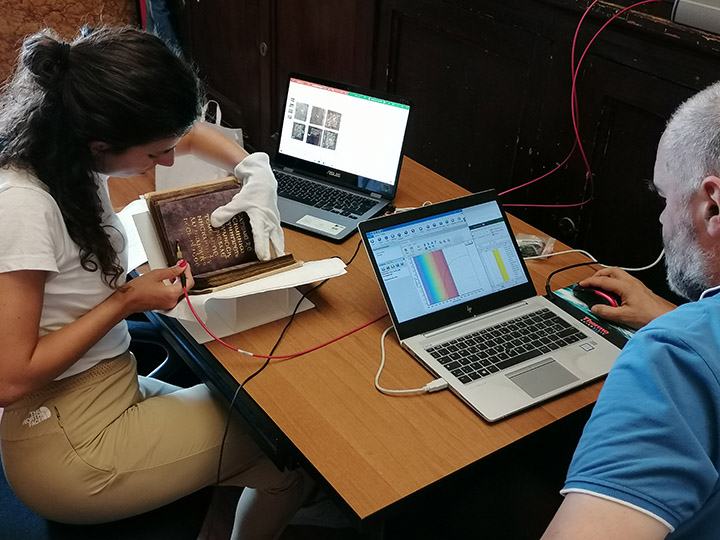
The Zeiss system (Carl Zeiss Spectroscopy GmbH, Jena, Germany) consists of a compact module that includes a light source and two spectroanalyzers in one chassis. The spectral resolution of the two spectroanalyzers—an MCS501 model operating in the 200–1000 nm range, and an MCS511 NIR 1.7 model operating in the 900–1700 nm range—was 0.8 and 6.0 nm/pixel, respectively. A 99% Spectralon diffuse reflectance standard was used to calibrate the spectroanalyzers.
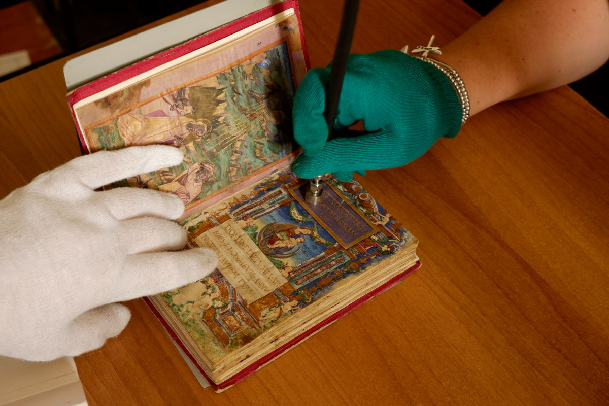
The Zeiss MCS 601 UV-NIR and MCS 611 NIR 2.2 WR spectroanalyzers are equipped with a dispersive grating and a linear detector of 1024 silicon and 256 indium gallium arsenide (InGaAs) photodiodes, respectively. The two spectroanalyzers are housed together in a compact and portable chassis, ideal for in situ analyses on different types of artworks. The radiation is provided by a voltage-stabilized 20 W halogen lamp (mod. CLH600) and is sent to and collected from the investigated area by means of quartz optical fiber bundles.
|
|
 |
 |
|
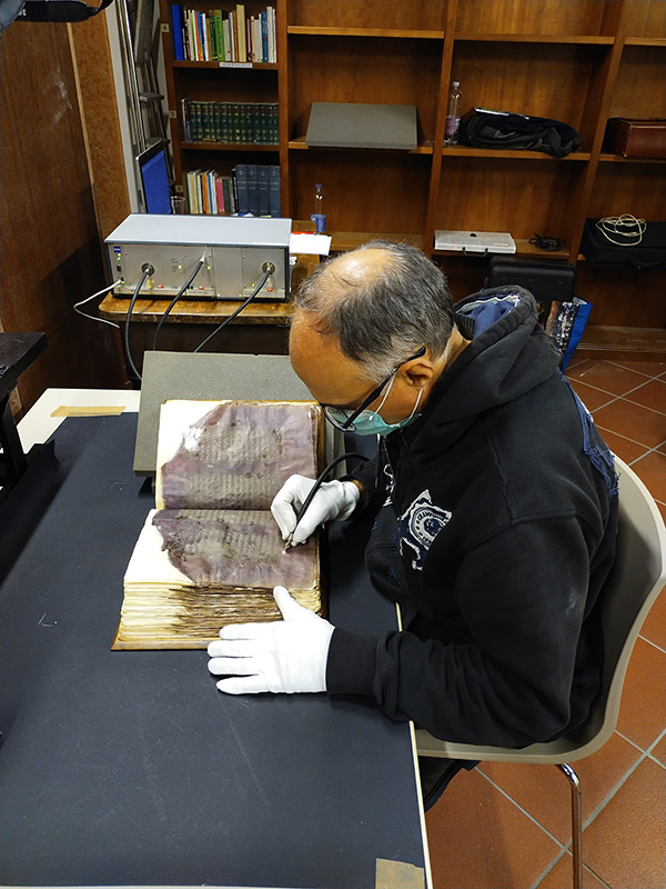
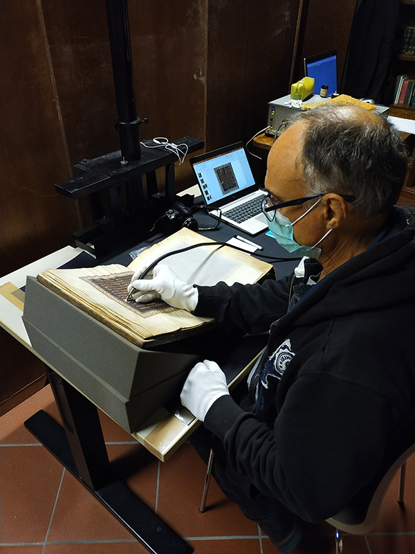
CNR-IFAC Zeiss system setup at the Biblioteca Capitolare di Verona and the Biblioteca Nazionale Vittorio Emanuele III of Naples. |
|
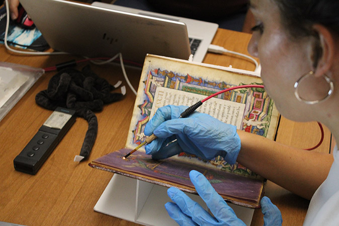
The second FORS device consisted of an Avantes (Apeldoorn, The Netherlands) spectrophotometer, model AvaSpec-ULS2048XL-USB2, and an AvaLight-HAL-S-IND tungsten-halogen source, both connected to a probe via an FCR-7UV200-2-1.5x100 branched optical fiber. The probe, with 45°/45° geometry to exclude the specular component, illuminated the surface at an angle of 45° and collected the backscattered radiation on the same axis. The sensitivity range of the spectrophotometer is between 200 and 1160 nm; depending on the characteristics of the monochromator (50 µm slit, grating with 300 lines/mm) and the detector (2048 pixels), a resolution of 2.4 nm FWHM is achieved.
The reflectance spectra were measured against a WS-2 standard (Avantes), which is guaranteed to be a 98% diffuser over the entire measurement range considered. The investigated area on the sample had a diameter of 1.5 mm. In all measurements, the distance between probe and sample was kept constant at 1 mm (corresponding to the focal length). A digital micro-camera connected to the PC via USB made it possible to view the investigated area. The instrumental conditions were as follows: 10 ms integration time, 100 acquisitions for a total of 1.0 s for each spectrum. The system was managed using the dedicated AvaSoft v. 8 software under Windows 7.
 |
 |
|
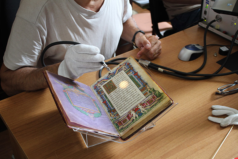
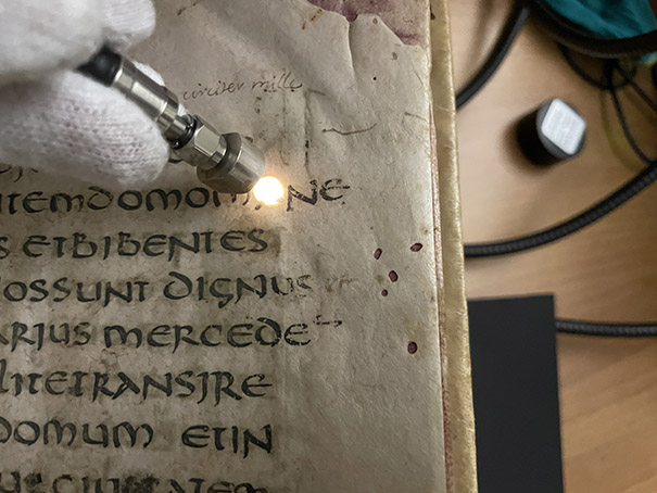
UPO Avantes system setup at the Biblioteca Nazionale Vittorio Emanuele III of Naples. |
|
References
- Aceto, M., Idone, A., Agostino, A., Fenoglio, G., Gulmini, M., Baraldi, P. and Crivello, F., 2014. Non-invasive investigation on a VI century purple codex from Brescia, Italy. Spectrochimica Acta Part A: Molecular and Biomolecular Spectroscopy, 117, pp.34-41.
- Aceto, M., Agostino, A., Fenoglio, G., Idone, A., Gulmini, M., Picollo, M., Ricciardi, P. and Delaney, J.K., 2014. Characterisation of colourants on illuminated manuscripts by portable fibre optic UV-visible-NIR reflectance spectrophotometry. Analytical Methods, 6(5), pp.1488-1500.
- Aceto, M., Calà, E., Agostino, A., Fenoglio, G., Idone, A., Porter, C. and Gulmini, M., 2017. On the identification of folium and orchil on illuminated manuscripts. Spectrochimica Acta Part A: Molecular and Biomolecular Spectroscopy, 171, pp.461-469.
- Aceto, M., Calà, E., Agostino, A., Fenoglio, G., Gulmini, M., Idone, A., Porter, C., Hofmann, C., Rabitsch, S., Denoël, C. and Förstel, C., 2019. Mythic dyes or mythic colour? New insight into the use of purple dyes on codices. Spectrochimica Acta Part A: Molecular and Biomolecular Spectroscopy, 215, pp.133-141.
- Cucci, C., Bracci, S., Casini, A., Innocenti, S., Picollo, M., Stefani, L., Rao, I.G. and Scudieri, M., 2018. The illuminated manuscript Corale 43 and its attribution to Beato Angelico: Non-invasive analysis by FORS, XRF and hyperspectral imaging techniques. Microchemical Journal, 138, pp.45-57.
- Picollo, M., Aceto, M. and Vitorino, T., 2019. UV-Vis spectroscopy. Physical Sciences Reviews, 4(4), p.20180008.
Keywords: FORS